Diagnostic Imaging
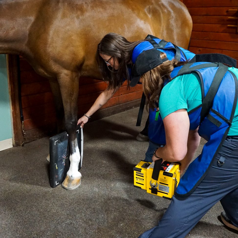
Radiographs are used to look at the bones of the musculoskeletal unit of the horse. The use of digital radiography provides a very crisp, detailed image instantly, allowing us to make decisions quickly. The images can then be sent through email, making them available to our clients, referring veterinarians and other clinicians worldwide at any time. Our portable unit allows us to take radiographs on farm or at horse shows and have the images instantly available for evaluation.
Balance Films can also be taken for your farrier to assist in properly shoeing your horse. The veterinarians at NWEP often work closely with farriers to find the best shoeing option for each horse.
Diagnostic ultrasonography is used extensively for the evaluation of tendons and ligaments to identify and monitor soft tissue injuries. The basic objective of an ultrasonographic evaluation is to characterize the size, shape, echogenicity (brightness) and fiber pattern of soft tissue structures in the region of interest. We can also use ultrasound to evaluate the bony surfaces of regions that may not be able to be imaged with other imaging, such as the pelvis. These findings are considered carefully in conjunction with the clinical examination to determine the significance of the injury. At NWEP we are fortunate to have a large ultrasound console to perform the highest quality ultrasound exams in the clinic. We also have the ability to perform complete exams in the field with our digital portable ultrasound unit.
MRI (Magnetic Resonance Imaging) gives us detailed pictures of your horse’s bones and soft tissues, making it easier to find and diagnose problems. It’s especially useful when other methods like X-rays or ultrasounds can’t clearly show what’s wrong. MRI can capture hundreds of images in one session, helping us see both bone and soft tissue issues at the same time.
At Northwest Equine Performance, we have a standing MRI system, which means your horse can remain standing during the scan, avoiding the risks of general anesthesia. We simply sedate your horse, and then scan the legs while they’re bearing weight—giving us a better idea of what’s causing any lameness or pain.
We are the only facility in Oregon and Washington with a standing MRI. This exam is safe and precise, allowing us to get a more accurate diagnosis, especially for issues like navicular disease or soft tissue injuries in the hoof. With MRI, we can identify problems early, which means faster treatment and a better outcome for your horse.
Upper Airway Endoscopy is performed to evaluate the upper airway nasal passages for structural and functional abnormalities.
Nuclear scintigraphy, or “bone scan,” is a technology borrowed from human medicine. A safe, low-dose radioactive material is injected into the horse’s vein. This material naturally collects in areas of the bone that are inflamed or injured. The bone scan detects the amount of material in the bone and creates an image of the area being examined.
A bone scan is useful for horses with performance issues or when lameness is hard to pinpoint because the horse doesn’t always show clear signs of lameness.
One of the main advantages of a bone scan is its high sensitivity, which can show problem areas that might look normal on an X-ray. Another benefit is that almost every bone in the horse’s body can be imaged without anesthesia, which helps in diagnosing issues in areas like the upper limbs, head, neck, back, and pelvis.
The horse will need to stay at the clinic for at least 24 hours after the injection because it will be temporarily radioactive. Horses should arrive early on the day of the scan, and the actual scan starts 2 hours after the injection. Scanning begins with the legs and moves to other areas like the back and neck. Any necessary follow-up is done the next day.
Gastroscopy is a diagnostic procedure used to examine your horse’s stomach, primarily for gastric ulcers. To prepare, horses must fast for 14-16 hours prior to scoping. Drop-off options include the day before or the morning of the procedure. Post-scope, horses can resume feeding once they’ve recovered from sedation. For more details or to schedule, contact our office.
Examinations
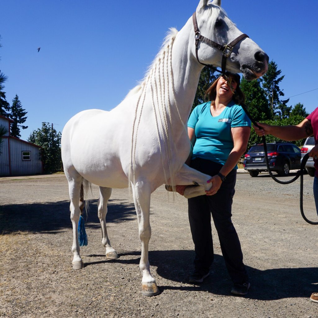
This hands-on evaluation of the horse is the cornerstone of the practice. Starting in the horse’s stall, we listen to the horse’s history and perform a thorough exam of the whole horse, focusing on various structures depending on the presenting complaint. The horse is then taken out for the moving part of the exam. Each horse is lunged on soft and hard surfaces. There is also a covered outdoor arena, so if your horse’s lameness is better viewed under saddle please bring your tack and a helmet. Flexion tests are then performed. We evaluate the horse’s response to flexions both passively (how they feel while we are performing the flexion) and actively (how they move after the flexion is done), Once the soundness evaluation is completed, a plan for further diagnostics or treatment is developed with owner/ rider/ trainer involvement.
A pre-purchase exam (PPE) is an overall evaluation of the horse’s current health, fitness and soundness at that time. There are many factors that go into a PPE pending the horse’s history, clinical concerns post exam and specific buyer concerns. Radiographs are often done, a complete set or just regions of concern can be imaged pending the buyer’s preference. Blood work is also commonly done, most often a Coggins (EIA) test. Other options include a drug screen, and/or a general chemistry / CBC to evaluate organ function and blood cell values to have a baseline on the horse.
Regenerative Therapy
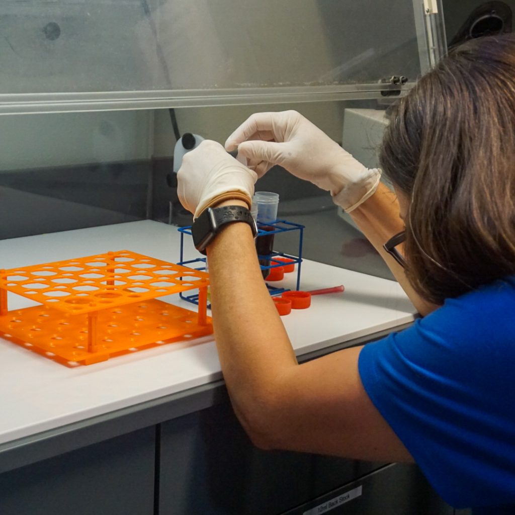
PRP therapy involves injecting a concentrated amount of platelets from the horse’s own blood into an injured area. Platelets contain growth factors and anti-inflammatory proteins that help speed up the healing process. The veterinarian takes a sample of the horse’s blood, processes it to isolate the platelet-rich portion, and then injects it into or around the injured spot.
PRP is often used to treat tendon and ligament injuries, as well as joint problems. In some cases, a shock wave treatment is done before the injection to enhance the therapy’s effects.
Shockwave therapy helps speed up the healing of soft tissue and bone injuries in horses. Despite the name, shockwaves aren’t electrical—they’re powerful sound waves that carry energy to deep areas of the body. This therapy acts like a controlled re-injury, reminding the body there’s a problem, which triggers new cell growth and improves blood flow to help with healing.
Injuries that respond well to shockwave therapy include arthritis (like low hock arthritis), ligament-related bone injuries (such as high suspensory injuries), soft tissue injuries, and back or pelvis issues (like kissing spines). Shockwave therapy can help injuries heal faster and stronger than with rest alone.
The treatment is quick, lasting about 10-15 minutes. The horse is sedated, and the vet selects the right settings based on the injury. Once the sedation wears off, the horse can go home. Shockwave therapy is often combined with PRP therapy, typically done before the PRP injection.
Horses are generally treated 2-3 times with about 14 days between sessions. We also offer on-farm shockwave therapy with our portable unit.
IRAP therapy is an anti-inflammatory treatment that helps prevent further tissue damage by blocking interleukin-1, a substance that causes inflammation during an injury. To create IRAP, we take a sample of the horse’s blood and mix it with special glass beads that boost the production of IRAP. The blood is then processed, and the IRAP is collected in syringes for immediate use or stored for future treatments.
IRAP is especially helpful for treating joint inflammation and arthritis, particularly in cases where other joint therapies are no longer effective.
A2M therapy is a new treatment for joint inflammation. A2M is an enzyme naturally found in the horse’s blood that helps protect cartilage by stopping its breakdown. It’s used in injured or inflamed joints to prevent further damage.
The procedure is done on an outpatient basis. The horse’s blood is drawn, processed, and filtered to create the A2M injections. Depending on how many joints need treatment, some A2M can be frozen for future use.
RenoVo is a treatment made from equine amniotic tissue, which has natural anti-inflammatory, anti-microbial, and healing properties. This tissue contains important proteins, growth factors, and cytokines that help with tissue repair.
RenoVo is used for soft tissue injuries that haven’t responded to other treatments, such as deep digital flexor tendonitis or chronic suspensory branch desmitis.
The FP4 system is a powerful therapeutic laser, much stronger than traditional laser therapy. Each treatment is customized for the patient and their specific injury. The FP4 uses four different laser wavelengths at once, allowing it to reach deep tissues without damaging the skin.
The laser’s wavelength is what determines how deep it can go and how well it’s absorbed by cells. Proven benefits of FP4 laser therapy include pain relief, reduced inflammation, better circulation, less swelling, reduced scar tissue, and the regeneration of healthy tissue. It can be used for both new and long-term injuries.
P3 therapy is an electromagnetic treatment used to help equine athletes recover from injuries. It works by increasing circulation in the treated area, which helps bring in healing factors and remove waste products. P3 therapy is great for pain relief, especially in horses with neck, back, or sacroiliac issues, but it can be used for any injury.
Our P3 machine is available for in-clinic treatments or for rental, so you can use it at your barn or even at a horse show. Call the office for more information!
Mesotherapy is a treatment that helps relieve pain by resetting the sensory nerves in the middle layer of the skin (mesoderm). Dr. Revenaugh first learned about this technique in 1992 while working at a veterinary clinic in Germany. Mesotherapy originated in France in 1952 and is commonly used in human medicine worldwide.
The treatment involves several small injections with fine needles that go just 4-6mm deep into the skin. After treatment, small bumps may appear on the skin but will disappear within a few hours as the injections are absorbed. The number of treatments and the type of products injected depend on the condition and how long it has been present.
Stem cell therapy involves injecting stem cells from bone marrow into an injury or through an IV. The bone marrow is collected in-clinic, usually from the horse’s sternum, and then sent to a lab to grow more stem cells, which takes 7-14 days. Once ready, the stem cells are injected into the injured area.
This therapy is mainly used for treating soft tissue injuries, and research is also being conducted on its use for treating laminitis in horses.
Amniotic Membrane based Biologic
A synthetic Polyacrylamide Hydrogel used in horses to provide improved joint lubrication.
Reduction of swelling and inflammation. Reduces muscle spasms, stiffness, and pain. Acceleration in recovery.